Dix choses que vous maîtriserez une fois formé à l’échographie clinique
L'échographie clinique est devenue un élément essentiel de l'examen physique, étant largement reconnue par la communauté médicale comme une technique d'imagerie médicale non irradiante et non invasive. Cette technique utilise des ondes sonores pour permettre une visualisation en temps réel des structures internes du corps, permettant ainsi un diagnostic d’orientation rapide et précis.
La formation à l'échographie clinique est devenue une pierre angulaire pour renforcer les capacités d’évaluation clinique du praticien. L’échographie vous permet d'améliorer la précision de vos diagnostics, de réduire le nombre d'examens complémentaires inutiles et d'optimiser la prise en charge des patients.
En 2022, The Ultrasound Journal a publié des recommandations sur les fondamentaux de l’échographie clinique que tout professionnel doit maîtriser à la fin de sa formation. Ces recommandations couvrent dix points clés, allant de la compréhension des principes physiques de l'échographie à l'intégration des résultats de l'échographie dans le processus de décision clinique. En voici notre récapitulatif :
Connaître l’écho-anatomie des différentes parties du corps
La pratique de l'échographie offre une compréhension plus concrète et précise de l'anatomie en permettant une visualisation directe et dynamique des structures anatomiques du corps humain, contrairement aux modèles anatomiques. Elle permet également de mieux comprendre les relations spatiales entre les différentes structures et d'apprécier les mouvements et les changements physiologiques. La pratique régulière de l'échographie aide à développer une compétence dans l'identification et la reconnaissance des structures anatomiques normales et anormales, ainsi que dans l'interprétation des images échographiques. Cette compétence est particulièrement utile dans les situations cliniques où une évaluation rapide et précise de l'anatomie est nécessaire pour poser un diagnostic et prendre des décisions thérapeutiques. De plus, la pratique de l'échographie favorise une approche plus globale de l'anatomie en intégrant les connaissances anatomiques avec les connaissances cliniques et physiologiques.
Maîtriser l'acquisition d'images
La pratique régulière de l'échographie clinique vous permet de développer une familiarité avec les différents types de sondes et les différents réglages de l'appareil d'échographie, ce qui vous permettra, au fur et mesure, d’optimiser la qualité des images acquises. Avec le temps, vous développerez une compréhension approfondie des principes fondamentaux de l'imagerie échographique, tels que la façon dont les ondes sonores interagissent avec les tissus du corps et comment les différents réglages de l'appareil affectent l'image produite. En visualisant et en interprétant régulièrement des images échographiques, vous développerez une solide capacité d’identification des structures anatomiques normales et anormales, ainsi qu’une aisance dans la reconnaissance des artefacts et des limitations de l'imagerie échographique.
Connaître l'examen FAST, pierre angulaire du POCUS en traumatologie
L'examen FAST (Focused Assessment with Sonography for Trauma) est une évaluation échographique rapide et ciblée utilisée pour détecter la présence de liquide libre dans la cavité abdominale ou péritonéale chez les patients traumatisés. Cette technique a été développée pour aider à détecter rapidement les hémorragies internes chez les patients présentant des signes vitaux instables après un traumatisme.A l’aide de votre sonde, vous visualisez quatre zones spécifiques du corps : la région sous-xiphoïdienne, la région sus-pubienne, les espaces latéraux entre le foie et le rein droit, et les espaces latéraux entre la rate et le rein gauche. L'objectif est de détecter la présence de liquide libre dans ces zones, ce qui peut indiquer une hémorragie interne.Il est important car il peut fournir des informations rapides et précieuses pour aider à guider la prise en charge de votre patient. En particulier, l'examen FAST peut vous aider à identifier les patients qui ont besoin d'une intervention chirurgicale urgente pour contrôler une hémorragie interne. Il peut également aider à éviter des interventions inutiles chez les patients qui n'ont pas d'hémorragie interne significative.
Comprendre les indications de l'échographie pulmonaire
L'échographie pulmonaire, longtemps considérée comme peu utile en raison de la présence d'air et des côtés, s'avère précieuse pour évaluer diverses pathologies respiratoires, telles que le pneumothorax, l'épanchement pleural et les pneumopathies infectieuses. L'interface pulmonaire offre des informations cruciales pour la prise en charge des pathologies pleurales, et l'échographie est plus adaptée que la radiographie du thorax pour le diagnostic de l’épanchement pleural réactionnel à une pneumonie et le suivi de l'abondance de la pleurésie.L'échographie est également un choix plus intéressant que la radiographie pour le diagnostic du pneumothorax résiduel après drainage ou du pneumothorax iatrogène secondaire aux gestes thoraciques. Dans le cas des pneumonies, les structures tissulaires ou hydriques visibles à la surface du poumon peuvent être détectées par échographie, qui a démontré une sensibilité supérieure à la radiographie pour la détection de pneumonie aux urgences.
Évaluer l’hémodynamique, le cœur et les vaisseaux
L'échographie cardiaque permet de visualiser le cœur en mouvement, d'évaluer la taille des cavités cardiaques, l'épaisseur et le mouvement des parois, ainsi que la fonction des valves cardiaques. Elle fournit également des informations sur la fraction d'éjection, un indicateur clé de la fonction cardiaque.L'échographie est aussi utilisée pour évaluer la compliance de la veine cave, un indicateur de la pression sanguine centrale et de la volémie, en mesurant le diamètre de la veine cave inférieure et ses changements pendant le cycle respiratoire. De plus, l'échographie permet d'évaluer la compressibilité des veines des membres inférieurs, une évaluation souvent utilisée dans le diagnostic de la thrombose veineuse profonde (TVP), en détectant si une veine se comprime complètement sous la pression de la sonde d'échographie ou non.
Appréhender les limites de l’échographie clinique
Les appareils d'échographie portables utilisés pour l’échographie clinique peuvent avoir une qualité d'image inférieure à celle des appareils d'échographie de grande taille, ce qui peut rendre la détection de certaines pathologies plus difficile. De plus, l'échographie clinique est limitée à des questions cliniques spécifiques et ne peut pas remplacer des examens d'imagerie plus approfondis tels qu'une échographie complète, une tomodensitométrie ou une IRM.L'échographie clinique est également limitée par la capacité de l'opérateur à visualiser les structures anatomiques, certaines zones du corps étant difficiles à visualiser en raison de la présence d'air, d'os ou de tissu adipeux. De plus, contrairement aux examens d'imagerie complémentaires, l’échographie clinique peut varier considérablement en fonction de l'opérateur et de la situation clinique. Il est donc important pour les professionnels de la santé de comprendre ces limites et d'utiliser l’échographie clinique de manière appropriée, en faisant appel à des examens complémentaires d'imagerie plus approfondis si nécessaire.
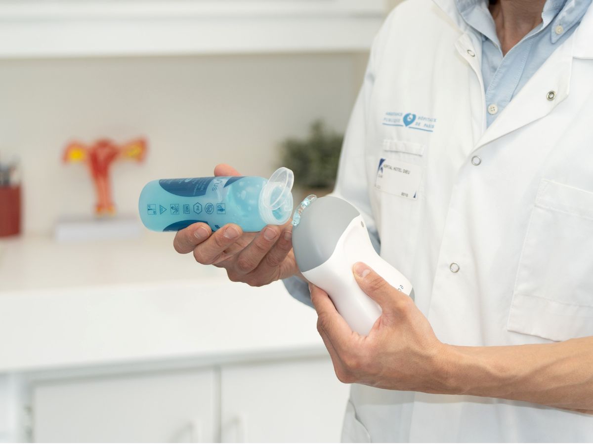
Désinfecter correctement la sonde et respecter les protocoles de stérilité
La désinfection correcte d'une sonde d'échographie est cruciale pour prévenir la transmission d'infections. Il est important de suivre les directives spécifiques de l'établissement de santé et du fabricant de l'équipement. Après chaque utilisation, essuyez la sonde avec une serviette douce pour enlever tout résidu de gel, puis désinfectez-la avec un désinfectant approuvé pour une utilisation avec des sondes d'échographie, tel que ceux à base d'alcool, de chlore ou d'iode. Suivez les instructions du fabricant concernant le temps de contact nécessaire pour une désinfection efficace. Inspectez la sonde pour détecter tout dommage ou usure et rangez-la dans un endroit propre et sec lorsqu'elle n'est pas utilisée.
Intégrer l’échographie dans votre processus de décision clinique
Pour intégrer efficacement l'échographie clinique dans votre prise en charge, une approche réfléchie et systématique est nécessaire. Tout d'abord, identifiez les situations cliniques où l'échographie pourrait fournir des informations utiles, telles que l'évaluation initiale des patients, le suivi de l'évolution d'une maladie ou la guidance de procédures. Ensuite, intégrez les résultats de l'examen dans le contexte clinique global du patient, en prenant en compte l'anamnèse, l'examen physique, les résultats de laboratoire et autres examens d'imagerie. Enfin, utilisez les résultats de l'échographie pour éclairer votre prise de décision clinique, que ce soit pour confirmer ou exclure un diagnostic, décider d'un traitement ou déterminer si des examens supplémentaires sont nécessaires.
Se tenir informé.e des dernières avancées
L'échographie clinique est un domaine en constante évolution, avec de nouvelles technologies, techniques et applications émergentes. En tant que professionnel de santé, il est important de se tenir informé.e de ces avancées pour offrir les meilleurs soins à vos patients. Participez régulièrement à des cours, des ateliers ou des conférences sur l'échographie clinique, lisez régulièrement des articles de revues scientifiques.Nous vous conseillons également d'adhérer à des sociétés savantes dans le domaine de l'échographie clinique. Ces organisations offrent souvent des ressources éducatives, des opportunités de réseautage et des mises à jour régulières sur les dernières avancées dans le domaine.
Développer un domaine de pratique ciblé
En vous concentrant sur un domaine spécifique de l'échographie clinique, vous pouvez développer une expertise approfondie et améliorer vos compétences dans ce domaine. Cela peut conduire à des diagnostics plus précis et à une prise en charge plus efficace de vos patients.En étant capable d'effectuer et d'interpréter des examens échographiques dans un domaine spécifique, vous pouvez également réduire le besoin de référer inutilement vos patients à d'autres spécialistes, ce qui peut améliorer la continuité des soins.Développer un domaine de pratique clé en échographie clinique peut également vous ouvrir des opportunités de recherche et d'enseignement. Vous pourrez contribuer à l'avancement du domaine en menant des recherches, en enseignant à d'autres professionnels de la santé ou en publiant des articles dans des revues médicales.
Conclusion
En conclusion, la formation à l'échographie clinique offre de nombreux avantages pour les professionnels de la santé. Elle permet de maîtriser l'écho-anatomie, l'acquisition d'images, l'examen FAST en traumatologie, les indications de l'échographie pulmonaire, l'évaluation de l'hémodynamique, du cœur et des vaisseaux, ainsi que la désinfection correcte de la sonde et le respect des protocoles de stérilité. De plus, la compréhension des limites de l'échographie clinique et l'intégration de cette technique dans le processus de décision clinique sont essentielles pour une utilisation efficace et sûre. Enfin, le développement d'un domaine de pratique ciblé et la participation à des activités de formation continue permettent de rester informé.e des dernières avancées dans ce domaine en constante évolution.
Formez-vous avec la sonde echOpen !
Pour en lire plus: Hoppmann, Richard A., et al. "International consensus conference recommendations on ultrasound education for undergraduate medical students." The Ultrasound Journal 14.1 (2022): 31.